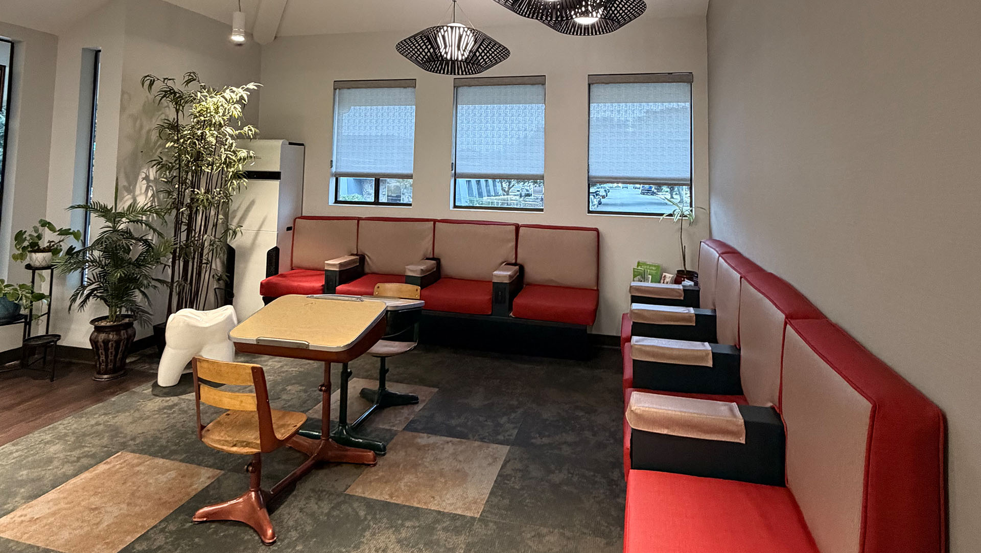
At Suezaki Family Dentistry, preventive care is the foundation of long-term oral health. One of the most important ways to maintain a healthy smile is through regular oral exams. By scheduling routine dental exams in San Jose, patients can stay ahead of potential concerns and receive personalized care designed to protect their teeth and gums for years to come.
Oral exams allow our team to monitor changes in your mouth, detect issues early, and recommend preventive treatments when needed. Rather than waiting for pain or visible problems to appear, routine check-ups in San Jose give us the opportunity to evaluate your overall oral health and help you maintain a confident, healthy smile.
Each dental exam combines a detailed visual evaluation, a review of your health history, and diagnostic imaging when necessary. This comprehensive approach allows us to build a clear understanding of your oral health and create a treatment plan that reflects your specific needs and goals.
Oral exams play an essential role in maintaining long-term dental health for both new and returning patients. Whether you are visiting our office for the first time or coming in for a routine check-up in San Jose, each exam allows our team to evaluate your current oral health, identify potential concerns, and ensure your care plan continues to meet your needs.
For new patients, the first visit includes a comprehensive oral exam designed to establish a clear baseline for your dental health. Oral exams for new patients in San Jose allow us to review your medical and dental history, understand any past treatments, and identify factors that may influence your oral health moving forward. This information helps us tailor preventive care and treatment recommendations specifically to you.
Returning patients benefit from regular dental exams in San Jose because these visits allow us to track changes in oral health over time. Even small developments can be detected early during routine check-ups, helping prevent minor concerns from progressing into more complex problems.
Each exam begins with a conversation about your health history, medications, and any dental concerns you may have noticed since your last visit. Certain medical conditions, lifestyle habits, and medications can affect oral health, so this discussion helps our team better understand possible risk factors and address them proactively.
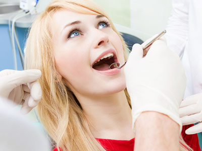
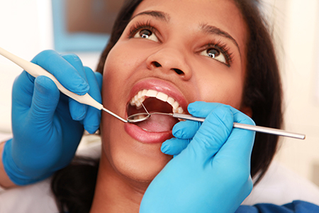
During the clinical portion of the oral exam in San Jose, we carefully evaluate your teeth, gums, and surrounding tissues. Our team checks for signs of cavities, gum inflammation, enamel wear, and other conditions that may require attention. We also examine how your teeth come together and assess the jaw joints for signs of strain or dysfunction related to the temporomandibular joint, often referred to as the TMJ.
If we observe signs of teeth grinding or clenching, we may recommend protective solutions that can reduce stress on your teeth and help prevent long-term damage. Addressing these concerns early is an important part of maintaining strong oral health.
In some cases, diagnostic imaging may be recommended to support the clinical exam. Dental X-rays and digital scans allow us to detect issues that cannot always be seen during a visual inspection, such as cavities between teeth, bone loss, or infections beneath the gumline.
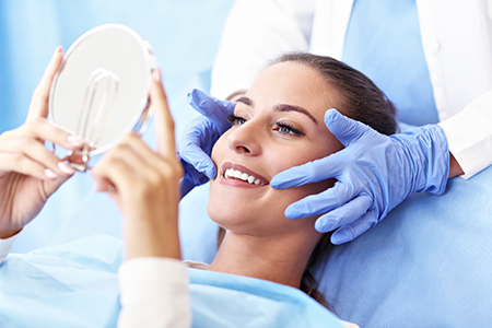
Once the dental exam is complete, our team will review the findings with you and discuss any recommendations for treatment or preventive care. We will also provide guidance on maintaining healthy habits at home so you can continue protecting your smile between routine check-ups in San Jose.
Your first visit includes a comprehensive evaluation designed to establish a clear baseline for your oral health. Whether you are scheduling oral exams for new patients in San Jose or returning to dental care after some time away, this appointment allows our team to gather important information and develop a preventive plan tailored to your needs. The visit typically begins with a discussion of your medical and dental history, including medications, past treatments, and any symptoms or concerns you may be experiencing. This conversation helps guide your dental exam in San Jose so we can focus on factors that may influence your oral health now and in the future.
During the clinical portion of the oral exam, we carefully evaluate your teeth, gums, and surrounding oral tissues. Our team checks for early signs of tooth decay, gum inflammation, enamel wear, and other conditions that could affect your smile. A periodontal assessment is also performed to evaluate the health of the gums and supporting structures around the teeth. As part of routine oral exams in San Jose, we also conduct an oral cancer screening and assess how your teeth come together when you bite. The temporomandibular joint, or TMJ, is examined as well to identify signs of strain, restricted movement, or discomfort that could be related to jaw function or habits such as teeth grinding.
Diagnostic imaging may be recommended if additional information is needed to complete the evaluation. Dental X-rays and digital scans help reveal issues that are not always visible during a visual exam, including cavities between teeth, bone loss, or infections beneath the gumline. Once your dental exam in San Jose is complete, our team will review the findings with you, answer any questions you may have, and discuss any recommended follow-up care. We will also provide practical guidance to help you maintain strong oral health between routine check-ups in San Jose.
Many patients are surprised to learn that oral exams can reveal much more than cavities or gum disease. A dental exam in San Jose can also provide valuable insight into your overall health.
The mouth often reflects early signs of systemic health conditions. Changes in gum tissue, persistent sores, or unusual lesions may signal underlying health concerns that deserve further evaluation. During your routine check-up in San Jose, our team carefully examines soft tissues, lymph nodes, and surrounding structures to identify abnormalities that may require attention.
Oral cancer screening is a standard component of our exams. Early detection plays a critical role in successful treatment, and routine screenings help ensure that any suspicious changes are identified as early as possible.
Oral health is also closely connected to other aspects of physical wellness. For example, chronic gum inflammation has been linked to complications associated with diabetes and cardiovascular health. In addition, certain medications can reduce saliva production, which increases the risk of tooth decay and oral infections.
By monitoring these factors during oral exams in San Jose, we can help patients better understand how their oral health relates to their overall well-being. Our team may also provide recommendations on lifestyle habits, hydration, oral hygiene routines, and other steps that support both dental and systemic health.
During each routine check-up in San Jose, we also pay attention to symptoms that patients sometimes overlook, such as persistent bad breath, changes in taste, or tooth sensitivity. While these concerns may seem minor, they can sometimes indicate underlying issues that benefit from early treatment.
Preventive dentistry relies on consistency. Regular dental exams in San Jose allow our team to track changes in your oral health over time and address problems before they become more complex.
Most patients benefit from scheduling a routine check-up in San Jose approximately every six months. These visits typically include a professional dental cleaning along with a comprehensive exam. Cleanings remove plaque, tartar, and bacterial buildup that cannot always be eliminated through brushing and flossing alone.
By removing these deposits, professional cleanings help reduce the risk of cavities and gum disease. They also leave your teeth feeling smooth and refreshed, making it easier to maintain good oral hygiene at home.
During routine oral exams in San Jose, we also provide personalized guidance to help patients improve their daily dental care habits. Even small adjustments to brushing techniques, flossing routines, or dietary choices can have a significant impact on long-term oral health.
Children benefit greatly from routine dental exams as well. For younger patients, these visits allow us to monitor the growth and development of the teeth and jaw. Early evaluations can help identify potential orthodontic concerns and guide families toward the appropriate timing for treatment if needed.
Preventive services such as fluoride treatments or dental sealants may also be recommended during certain stages of childhood development. These treatments add an extra layer of protection for teeth that may be more vulnerable to cavities.
By attending regular routine check-ups in San Jose, patients of all ages can maintain healthier smiles and reduce the likelihood of needing more extensive dental treatments later on.
Modern dental exams rely on advanced technology to improve diagnostic accuracy and provide patients with a clearer understanding of their oral health. Digital imaging plays an important role in many oral exams in San Jose because it allows our team to evaluate areas of the teeth and supporting structures that cannot be seen during a visual examination alone.
Digital X-rays and other imaging tools provide detailed views of tooth roots, bone levels, and the spaces between teeth. These images help dentists detect cavities that form between teeth, identify early bone loss, and uncover infections or abscesses that may develop beneath the gumline. By capturing this information, imaging supports a more complete and accurate dental exam in San Jose.
Compared with traditional film radiography, digital imaging offers several advantages for both patients and dental professionals. Images can be captured quickly, reducing the amount of time patients spend in the chair during a routine check-up in San Jose. Digital systems also use significantly less radiation while producing highly detailed images that appear instantly on a monitor. This allows our team to review results with patients in real time and explain any findings clearly.
Digital imaging also makes it easier to maintain accurate records of your oral health over time. By comparing images taken during different visits, we can monitor changes in your teeth and gums and evaluate how well preventive care or treatments are working.
Different types of dental images may be used depending on what your dentist is evaluating during your oral exam in San Jose. Bitewing X-rays are commonly taken during routine dental exams because they help detect cavities that develop between teeth, an area that is difficult to examine visually. Periapical images provide a detailed view of a single tooth from the crown down to the root and are useful for diagnosing infections, root problems, or changes in the surrounding bone.
In some cases, a full mouth series may be recommended to provide a comprehensive view of all teeth and supporting structures. Panoramic X-rays offer a broader perspective of the jaws and surrounding anatomy and can help identify impacted teeth, evaluate jaw development, or assess other structural concerns.
For more complex situations, advanced three-dimensional imaging may be recommended. Cone-beam computed tomography, commonly referred to as CBCT, creates a highly detailed 3D view of the teeth, bone, and surrounding structures. This technology is often used when planning treatments such as dental implants, oral surgery, or evaluating more complex dental conditions.
Whenever imaging is recommended as part of your dental exam in San Jose, our team will explain why it is needed and how it contributes to a more accurate diagnosis. By combining thorough clinical evaluation with modern imaging technology, we are able to deliver more precise care and develop treatment plans designed to protect your long-term oral health.
Consistent dental care is one of the most effective ways to protect your smile. Routine oral exams in San Jose help detect problems early, support preventive care, and provide patients with the guidance they need to maintain healthy teeth and gums.
At Suezaki Family Dentistry, our goal is to create a comfortable and informative experience for every patient. Whether you are visiting for a routine check-up in San Jose or scheduling oral exams for new patients in San Jose, our team is committed to providing thorough, personalized care.
By combining clinical expertise, modern technology, and patient education, we help individuals and families maintain strong oral health throughout every stage of life.
If you are due for a dental exam in San Jose or would like to schedule your first visit, we invite you to contact our office today to learn more about our comprehensive approach to preventive dental care.
A comprehensive oral exam combines a review of your medical and dental history with a thorough clinical evaluation of the teeth, gums, and surrounding soft tissues. The clinician performs a visual inspection and hands-on assessment to check for signs of decay, gum inflammation, abnormal lesions, and changes in tooth position or wear. This exam also includes a focused screening for oral cancer and an evaluation of the temporomandibular joint and bite relationship to identify functional problems.
When indicated, the exam is complemented by diagnostic imaging to reveal findings that are not visible to the naked eye, such as interproximal decay, root issues, or bone loss. After the assessment, the clinician reviews the findings with you, outlines recommended next steps, and creates a personalized prevention or treatment plan based on your risks and goals. The goal is to detect problems early and provide practical guidance to maintain or improve oral health.
Your initial oral exam is designed to establish a baseline and inform an individualized plan of care. The visit begins with a conversation about your medical history, medications, and any concerns so the exam can be tailored to your needs and risk factors. Clinicians then perform a systematic evaluation of teeth, gums, oral soft tissues, and jaw function, documenting any abnormalities and identifying areas that require monitoring.
If imaging is needed, the team will recommend appropriate films or digital scans and explain how those images will help with diagnosis and planning. You will receive a clear summary of findings at the end of the visit along with preventive recommendations and any suggested follow-up care. At Suezaki Family Dentistry we emphasize patient education so you leave understanding the rationale for any recommended steps.
Frequency for routine oral exams depends on individual risk factors, oral health status, and life stage rather than a one-size-fits-all interval. Many adults benefit from checkups and professional cleanings every six months to manage plaque, monitor small changes over time, and treat conditions in their earliest stages. Patients with a history of periodontal disease, frequent decay, certain medical conditions, or medications that affect saliva may require more frequent visits to control risk.
An oral exam can provide valuable clues about systemic health because the mouth often reflects conditions that affect the rest of the body. Signs like persistent gum inflammation, unusual oral lesions, dry mouth, or changes in tissue color and texture can be associated with diabetes, nutritional deficiencies, autoimmune conditions, or medication side effects. By documenting these findings and tracking them over time, dental clinicians can advise patients when medical follow-up or collaborative care with a physician is appropriate.
Addressing oral inflammation and infection can also support better systemic outcomes, for example by reducing inflammatory burden that may affect metabolic control. During an exam, the team discusses how lifestyle factors, medications, and chronic conditions influence oral risk and recommends practical steps patients can take at home to support both oral and general health. Early identification and coordination of care improve the chances of timely, effective treatment.
Oral cancer screening is an essential component of a thorough oral exam because early detection greatly improves treatment outcomes. The screening includes inspection and palpation of the lips, tongue, floor of mouth, cheeks, palate, and neck lymph nodes to identify persistent ulcers, white or red patches, lumps, or other suspicious changes. Clinicians document any abnormalities and may recommend closer monitoring, adjunctive screening tools, or referral for biopsy when warranted.
Patients are also counseled about risk factors such as tobacco use, heavy alcohol consumption, and HPV exposure, as well as signs to watch for between visits. Prompt evaluation of persistent or unexplained oral changes helps catch potentially serious conditions at an earlier, more treatable stage. Routine screening is a preventive measure that complements other diagnostic steps taken during the exam.
Digital imaging significantly improves diagnostic accuracy by revealing issues beneath the visible surface, such as interproximal decay, bone levels, and root problems. Compared with traditional film, digital radiography and three-dimensional scans capture images more quickly, involve less radiation exposure, and allow immediate review during the appointment. Clinicians use these images to inform diagnoses, communicate findings clearly to patients, and plan conservative, targeted treatments when needed.
Three-dimensional cone-beam computed tomography (CBCT) is used selectively for complex cases like implant planning, impacted teeth evaluation, or surgical assessment because it provides volumetric detail not available on two-dimensional films. The diagnostic team balances the need for detailed information against the principle of minimizing exposure, choosing the least invasive modality that will yield reliable results. Storing images digitally also enables easy comparison over time and secure sharing with specialists when collaborative care is required.
Different images serve different diagnostic purposes: bitewings are excellent for detecting decay between teeth, periapical views capture a whole tooth from crown to root and help evaluate roots and supporting bone, and full mouth series provide a comprehensive baseline when an in-depth survey is needed. Panoramic films offer a broad view of the jaws and are useful for assessing growth, impacted teeth, and overall development. Cephalometric images are specialized profile films commonly used in orthodontic assessment to analyze jaw relationships and facial proportions.
CBCT scans are reserved for cases where three-dimensional anatomy is critical, such as implant placement, evaluation of complex pathology, or planning surgical procedures. The care team will explain why a particular image is recommended and how it will affect diagnosis or treatment, ensuring you understand the clinical benefit before the image is taken. Using the right image for the clinical question improves safety and treatment predictability.
Periodontal evaluation includes measuring pocket depths around teeth, assessing gum inflammation and bleeding, and checking for recession or attachment loss that indicate tissue support has been compromised. The clinician examines the gums for color, contour, and texture, and evaluates mobility and bone support through clinical measures and appropriate imaging. These findings are recorded to establish a baseline and to monitor progression or improvement over time.
Based on the assessment, the team recommends preventive strategies such as enhanced home care instruction, more frequent professional cleanings, or nonsurgical periodontal therapy when indicated. Early intervention and consistent maintenance greatly reduce the risk of significant bone loss and tooth instability. Patients are guided on practical steps to reduce inflammation and maintain periodontal health between visits.
Evaluation of the temporomandibular joint (TMJ) includes observing jaw movement, listening for clicking or popping, checking for deviations on opening, and palpating muscles for tenderness or tightness. The clinician asks about symptoms such as facial pain, headaches, jaw locking, or tooth sensitivity that can be associated with bruxism (clenching and grinding). Clinical findings combined with a review of habits, stressors, and sleep history help determine whether the issue is primarily muscular, joint-related, or dental in origin.
Treatment recommendations can range from conservative approaches—like occlusal adjustments, behavioral strategies, night guards, and targeted muscle therapy—to referrals for specialized care when structural joint concerns are identified. The objective is to protect tooth structure, relieve discomfort, and restore comfortable function using the least invasive measures appropriate for the diagnosis. Ongoing monitoring during routine exams helps track response to therapy and detect any evolution of symptoms.
To schedule an oral exam, you can contact the office by phone or request an appointment online; the team will confirm details and let you know any preparatory steps to take. Bring a list of current medications, relevant medical records, and information about any medical conditions or recent surgeries so the clinician can consider these factors during the assessment. If you have dental insurance, bring your card or plan information to help the administrative team with claims processing and appointment paperwork.
Arrive with a brief list of concerns or symptoms you want to discuss so the visit addresses your priorities efficiently, and wear comfortable clothing for a relaxed experience. The staff in San Jose will ensure your records are secure and that you receive a clear explanation of findings and recommended next steps before you leave. If you have questions about what to expect, the team at Suezaki Family Dentistry can provide guidance when you call to schedule.
Absolutely. Most of our preventative cleanings take about 45 to 60 minutes. By choosing a dentist close to me right here in San Jose, you can swap a long commute for a refreshing scale and polish during your lunch hour, heading back to work with a brighter smile and zero "travel fatigue."