An intraoral camera is a compact, pen-sized imaging device designed specifically for dental use. It captures clear, full-color images of teeth, gums, and other oral structures and transmits them instantly to a monitor. Because the camera can reach locations that are difficult to view directly, it reveals details that might otherwise be missed during a visual exam alone.
Modern intraoral cameras produce high-resolution stills and video, giving clinicians a magnified, real-time view of small cracks, early decay, worn restorations, and soft-tissue changes. This level of detail helps the dental team establish an accurate baseline, track changes over time, and tailor care based on what is actually visible inside the mouth rather than relying solely on descriptions.
Although the device is small, its diagnostic value is large: the intraoral camera bridges the gap between clinical observation and the images stored in a patient’s record. Because images can be reviewed at any time, they become part of an ongoing conversation about oral health—one rooted in clear visual evidence rather than abstract explanation.
One of the clearest benefits of intraoral imaging is its power to improve communication between clinician and patient. When a patient can see an enlarged image of a tooth or an area of redness on a monitor, complex issues become easier to grasp. Visual evidence helps patients understand the condition, recommended treatments, and the reasons behind clinical decisions.
Sharing images in real time encourages collaboration; patients can ask targeted questions, and clinicians can point out exactly where a problem exists and why it matters. This shared view supports informed decision-making and reduces uncertainty, because the discussion is anchored to the same visual reference rather than memory or description alone.
For many patients, visualization reduces anxiety by demystifying the mouth’s condition. When the dental team takes time to explain what the image shows and what the possible next steps are, patients often feel more confident about their care plan and more invested in maintaining oral health.
Intraoral cameras are versatile diagnostic tools used across preventive, restorative, and periodontal care. Practitioners use them to detect early enamel breakdown, monitor margin integrity around crowns and fillings, identify cracks or fractures, and document the health of soft tissues. Because the camera can reveal subtle color and texture differences, it often highlights issues that merit closer inspection or further testing.
Beyond detection, intraoral images support monitoring. Photographs taken during routine visits create a visual timeline that makes it easier to compare changes over months or years. This is especially valuable for tracking lesion development, assessing the stability of restorations, and measuring the progression or improvement of gum conditions.
Images captured with the intraoral camera also serve as a clinical record. When a clinician documents an area of concern with clear photographs, subsequent appointments begin with a concrete reference point. That continuity strengthens diagnostic accuracy and allows for better-coordinated care among clinicians when multiple providers are involved.
Intraoral cameras integrate smoothly with modern digital dental systems. Captured images can be stored directly in the patient’s electronic chart, organized by date and specific tooth, and retrieved during future visits. This digital compatibility streamlines recordkeeping and improves the efficiency of clinical workflows without adding manual steps.
Images from the intraoral camera can also be shared with other dental specialists or laboratories when collaborative care is required. Because the photographs provide a clear, objective depiction of the oral condition, they help specialists understand the case quickly and contribute to smoother treatment planning and execution.
The camera’s output complements other digital tools—such as digital radiography, CBCT, and CAD/CAM systems—by offering surface-level photographic detail that radiographs do not capture. Together, these technologies create a comprehensive diagnostic picture that supports precise, conservative treatment decisions.
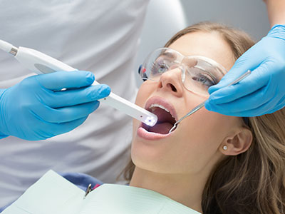
Undergoing an intraoral camera exam is straightforward and comfortable. During a routine checkup or targeted assessment, the clinician will gently guide the camera into the mouth to capture still images or short video clips. The process is noninvasive and typically adds only a few minutes to the appointment, while delivering a wealth of useful information.
Images are displayed on a chairside monitor so patients can view them as they are taken. Clinicians will often pause to explain what each image shows, point out areas of concern, and discuss why a certain observation might warrant treatment or closer monitoring. This step-by-step review makes recommendations easier to understand and helps patients participate in their care.
All images taken during the visit become part of the patient’s secure dental record. Clinicians follow privacy and recordkeeping standards to protect patient information while ensuring those images are available for future comparison, specialist consultations, or laboratory needs as appropriate.
At Suezaki Family Dentistry, intraoral cameras are one of the many tools we use to provide precise, transparent care. If you’d like to learn more about how intraoral imaging is used during exams or how it can help you better understand your oral health, please contact us for more information.
An intraoral camera is a small, pen-sized imaging device that captures high-resolution stills and short video of the teeth, gums and other oral structures. The clinician guides the camera inside the mouth while images are transmitted instantly to a chairside monitor, providing a magnified, full-color view of areas that are difficult to see with the naked eye. Illumination and close-up optics reveal surface details such as small cracks, early enamel breakdown and soft-tissue changes.
The images are saved directly to the patient’s digital chart for documentation and comparison over time. Because the camera offers real-time visual feedback, it supports immediate discussion of findings and treatment options. Many modern intraoral systems also allow clinicians to annotate or zoom images to highlight specific concerns during the visit.
Intraoral imaging helps patients see exactly what the clinician sees, which improves understanding of diagnoses and recommended care. Visual evidence makes it easier to grasp the location and extent of a problem, reducing uncertainty and enabling more informed questions and decisions. This transparency often increases patient comfort and trust because recommendations are based on clear photographic documentation.
Beyond communication, intraoral photos create a visual baseline to track changes over months or years, which is especially useful for monitoring restorations, lesions and gum conditions. The images also support continuity of care by providing objective records that can be reviewed at subsequent visits or by other members of the dental team. Overall, intraoral imaging contributes to more collaborative and evidence-based care.
An intraoral camera can reveal subtle surface changes that are difficult to observe directly, such as tiny fractures, early enamel demineralization and gaps around restoration margins. The magnification and lighting can highlight color and texture differences in soft tissue that may indicate inflammation or atypical lesions. These details help clinicians decide whether closer inspection, additional testing or preventive measures are warranted.
While the camera excels at surface-level visualization, it complements rather than replaces other diagnostic tools like radiographs and CBCT when deeper structures or bone involvement need evaluation. Images from the intraoral camera are often the first indicator that a targeted radiograph or periodontal assessment is necessary, which improves diagnostic accuracy and helps avoid missed problems.
Yes. Intraoral imaging is noninvasive and does not expose patients to radiation, making it a safe option for most people, including children and pregnant patients. The camera is small and designed for intraoral use, and clinicians typically cover the tip with a disposable sheath or use other infection-control measures to maintain hygiene. Most patients experience little or no discomfort, and the procedure usually adds only a few minutes to a routine appointment.
The practice follows standard sterilization and barrier protocols to protect patient safety and privacy when capturing and storing images. Because the camera provides immediate visual feedback, clinicians can pause and explain findings as images are taken, which many patients find reassuring. If a patient has a sensitive gag reflex or limited mouth opening, the clinician can adapt technique or use positioning aids to increase comfort.
During an intraoral camera exam the clinician will gently insert the camera into your mouth and capture a series of still images or short video clips of relevant areas. Images are displayed on a chairside monitor so you can view them as they are taken and follow along while the clinician explains what each image shows. The process is quick, typically noninvasive and usually only extends the appointment by a few minutes.
The clinician may take images of specific teeth, restoration margins, soft-tissue sites or other areas of concern and will save those photos in your digital record for future comparison. You are encouraged to ask questions while the images are reviewed, which supports shared decision-making and helps you understand recommended next steps. All images are stored according to privacy and recordkeeping standards.
Intraoral images become part of the secure electronic record and are organized by date and tooth or site to create a visual timeline of oral health. Clinicians use this documentation to compare current findings with past images, evaluate the progression or stability of conditions and verify the integrity of restorations over time. Clear photographic records improve diagnostic continuity and reduce ambiguity when reviewing a case at future visits.
When developing a treatment plan, images help clinicians explain options and illustrate why a particular approach is recommended, which supports informed consent. They also provide objective evidence that can be referenced when coordinating care with specialists or when laboratory communication is required. This integration with the patient record enhances both accuracy and efficiency in clinical workflows.
Yes. High-quality intraoral photographs can be securely shared with dental specialists and dental laboratories to facilitate collaborative case planning and prosthetic design. Clear images help receiving providers quickly understand the clinical situation, assess restorative margins, evaluate soft-tissue contours and recommend next steps without requiring redundant intraoral exams. Sharing images reduces miscommunication and speeds the planning process for multi-provider care.
When images are transmitted, the practice follows privacy and recordkeeping protocols to protect patient information and ensure secure exchange. Photos are typically paired with clinical notes, radiographs and digital impressions as needed to give specialists and labs a comprehensive view of the case. This coordinated approach supports more precise and predictable treatment outcomes.
An intraoral camera provides surface-level photographic detail that complements radiographic images, CBCT and CAD/CAM scans by showing color, texture and visible defects that X-rays cannot capture. While radiographs and CBCT reveal internal structures and bone relationships, intraoral photos document the external appearance of teeth and soft tissues, which is essential for aesthetic assessment and restorative planning. Together, these tools create a fuller diagnostic picture for conservative, targeted treatment.
In restorative workflows intraoral images can assist with shade selection, margin evaluation and communication with dental laboratories or milling centers. When used alongside digital impressions and CAD/CAM systems, camera images help ensure restorations fit and blend properly with surrounding tissues. This multimodal approach improves clinical decision-making and the overall quality of care.
Yes. Although intraoral cameras reveal excellent surface detail, they cannot substitute for radiographs or CBCT when bone-level assessment or detection of subsurface pathology is required. The camera does not show tooth roots, interproximal decay beneath contact points or bone loss in three dimensions, so additional imaging or diagnostic tests may be necessary to reach a comprehensive diagnosis. Clinicians use intraoral images as one component of a wider diagnostic strategy.
Additionally, some findings that appear suspicious on camera images will require further evaluation, biopsy or specialist referral to determine their nature. Intraoral imaging is a valuable screening and documentation tool, but definitive diagnosis often relies on a combination of visual, tactile, radiographic and histologic information. Your clinician will recommend the appropriate next steps when camera images indicate further assessment is warranted.
Yes. The office routinely uses intraoral cameras as part of comprehensive exams to improve visualization, record findings and help patients understand their oral health. Incorporating intraoral imaging into regular visits supports early detection of problems, clearer communication about recommended care and consistent documentation for comparison over time. The practice pairs these images with digital charts and radiographs to create a complete clinical record.
If you would like to see intraoral images during your appointment or learn how they are used in your own treatment planning, the team at Suezaki Family Dentistry will be happy to explain the process and review your photos. Viewing images together helps ensure that you understand any findings and feel confident in the recommended approach to care. Please ask your clinician at your next visit if you would like intraoral imaging included in your exam.