Digital radiography is the modern approach to dental X-rays that replaces traditional film with electronic sensors and computer processing. Instead of chemical development, images are captured on a sensor and immediately converted into high-resolution digital files. This shift from analog to digital fundamentally changes how clinicians view, manipulate, and store radiographic images, making the entire imaging process faster and more adaptable for patient care.
For patients, the most visible benefit is convenience: images appear on a monitor within seconds, allowing dentists to explain findings right away during the appointment. For clinicians, the clarity and manipulability of digital images enhance diagnostic accuracy—subtle differences in density and structure can be highlighted, magnified, or adjusted without retaking the image. That combination of speed and precision helps the dental team make confident recommendations on the same day.
At the office of Suezaki Family Dentistry, digital radiography is integrated into routine exams and treatment planning so that decisions are data-driven and easy to share with patients. By investing in up-to-date imaging tools, the practice aims to reduce uncertainty, shorten appointment times, and improve the overall patient experience without compromising diagnostic quality.
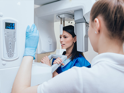
One of the defining advantages of digital radiography is how quickly images become available for review. The moment a sensor records an exposure, the image is transmitted to the computer where the clinician can view it, adjust contrast and brightness, and zoom in on areas of interest. This immediacy removes the delay associated with film processing and enables the dentist to identify issues—cavities, bone loss, or root structure—during the same visit.
Beyond speed, digital images often provide superior clarity. Software tools can enhance fine details, making it easier to detect early-stage problems that might be missed on traditional film. Image enhancement features also support patient education: when clinicians can point out and annotate findings in real time, patients gain a clearer understanding of their oral health and the rationale behind recommended treatments.
This combination of clarity and speed helps streamline diagnosis and care coordination. Whether planning restorative work or monitoring progressive conditions, having immediate access to high-quality images reduces guesswork and supports more precise clinical decisions that prioritize long-term oral health for every patient.
Digital radiography significantly reduces radiation exposure compared with conventional film X-rays. Because sensors are more sensitive to X-ray photons, images can be captured with lower doses while maintaining diagnostic quality. This is especially meaningful for patients who require frequent imaging—children, patients with ongoing restorative needs, and those undergoing multi-stage treatments benefit from lower cumulative exposure.
Safety is further enhanced by the ability to limit retakes. Digital sensors deliver predictable image quality, and immediate feedback lets clinicians confirm a successful capture before the patient leaves the chair. When a retake is necessary, adjustments can be made in real time to avoid repeat exposures. These practical safeguards contribute to a more patient-centered approach to radiography.
Practices using digital radiography also tend to follow contemporary radiographic protocols that evaluate imaging needs on a case-by-case basis. The objective is always to obtain the diagnostic information required to manage care effectively while keeping patient safety and radiation stewardship at the forefront.
Digital X-rays are more than a diagnostic snapshot; they are a foundation for treatment planning. Clear, manipulable images allow dentists to assess bone levels, visualize root anatomy, and plan restorations with greater precision. Whether preparing for crowns, evaluating a suspected root canal, or assessing periodontal health, digital images support clinical judgment and help foresee potential complications before treatment begins.
Integration with digital workflows means radiographic data can be combined with other technologies—such as digital impressions and in-office CAD/CAM systems—to create cohesive, predictable treatment pathways. For example, a digital radiograph can guide the placement and restoration of a crown, while intraoral scans capture surface detail. Together, these tools reduce manual steps and increase the likelihood of successful, efficient outcomes.
Because digital images can be annotated and archived, they also support long-term case tracking. Clinicians can compare sequential images to evaluate healing, monitor disease progression, or measure the stability of a restoration. This historical perspective is valuable for both routine maintenance and complex, multi-phase treatments.
Digital radiography simplifies image storage and retrieval by maintaining electronic files within the patient’s digital record. Rather than handling physical films, clinicians can access images from any authorized workstation in the office, view prior studies instantly, and include radiographs in a patient’s ongoing chart. This organized approach improves recordkeeping and reduces the risk of lost or degraded films over time.
Collaboration with specialists is also easier with digital images. Files can be shared securely with referral partners or consulting clinicians for second opinions, surgical planning, or interdisciplinary care coordination. When images are transmitted electronically, they arrive quickly and remain intact, preserving detail that’s critical for accurate assessment. Secure transfer protocols and practice policies ensure that patient privacy is protected during these exchanges.
For patients, the result is more seamless care: fewer trips to reassign films, faster consultations with specialists, and clearer explanations about treatment direction. From a practice perspective, efficient image management supports better communication, smoother referrals, and a higher standard of continuity in patient care.
Wrap-up: Digital radiography represents a meaningful advancement in dental imaging—combining speed, image quality, and safety to support accurate diagnoses and efficient treatment planning. By reducing radiation exposure, enabling immediate review, and simplifying secure storage and sharing, digital X-rays help clinicians deliver thoughtful, evidence-based care. If you’d like to learn more about how digital radiography is used during exams and treatment at our practice, please contact us for more information.
Digital radiography is the process of capturing dental X-ray images with electronic sensors and converting them into high-resolution digital files that can be viewed and manipulated on a computer. This technology replaces traditional film and chemical processing with instant image acquisition and software-based enhancement tools. The result is faster diagnosis, clearer visualization of structures, and more efficient communication between clinician and patient.
Digital radiography matters because it supports evidence-based decision making throughout care, from routine exams to complex restorative planning. Immediate access to images shortens appointment flow and enables clinicians to demonstrate findings in real time. For practices focused on modern, patient-centered care, digital imaging is a practical cornerstone of accurate and timely treatment.
Unlike film X-rays, digital radiography uses electronic sensors that capture images and transmit them instantly to a computer for review. There is no chemical development, which eliminates processing delays and the need to store physical films. Software tools also allow clinicians to enhance contrast, zoom, and annotate images without retaking exposures.
Digital images are easier to archive, retrieve, and share compared with analog film, which can degrade or be misplaced over time. The reproducibility and immediate feedback from sensors reduce the likelihood of repeat exposures and support more consistent imaging quality. These practical differences improve both clinical workflow and long-term recordkeeping.
Digital dental X-rays use much lower radiation doses than conventional film because modern sensors are more sensitive to X-rays, allowing diagnostically useful images with reduced exposure. For routine dental care, digital imaging is generally considered safe for adults and children when used according to accepted radiographic guidelines. Protective measures such as thyroid collars and lead aprons are used as appropriate to minimize exposure.
If pregnancy is confirmed or suspected, inform your dentist so imaging can be evaluated and scheduled with additional precautions or postponed when clinically appropriate. The clinician will weigh the diagnostic benefit against any potential risk and follow established protocols to protect the patient. For specific medical concerns, patients should consult their obstetric provider in addition to their dental team.
Digital radiography streamlines the appointment by producing images within seconds, allowing the dentist to review and explain findings during the same visit. This immediacy reduces idle time in the chair and helps patients understand their condition through on-screen visuals and annotations. Because sensors provide rapid feedback, clinicians can confirm image quality immediately and avoid unnecessary retakes.
Patients often appreciate the clearer explanations that digital images provide, which can improve informed consent and collaborative treatment planning. The reduced radiation dose and shorter imaging time also enhance comfort and safety, particularly for younger patients and those who require frequent monitoring. Overall, digital workflows contribute to a more transparent and efficient visit.
Digital radiographs provide detailed information about tooth structure, root anatomy, bone levels, and surrounding tissues that is essential for planning restorations, endodontic treatment, and periodontal therapy. High-resolution images allow clinicians to detect decay, fractures, or anatomical variations that influence the choice and timing of procedures. When combined with clinical examination and other diagnostics, radiographs support precise, predictable treatment pathways.
Integration with other digital tools, such as intraoral scans and CAD/CAM systems, creates a cohesive workflow for same-day restorations and complex rehabilitations. Radiographic data can be annotated and referenced during laboratory communication or surgical planning to reduce uncertainty and anticipate potential challenges. This combined approach improves accuracy and helps achieve consistent clinical outcomes.
Software tools for digital radiography enable clinicians to adjust brightness and contrast, apply filters, and zoom into areas of interest to reveal subtle changes that might be missed on film. These enhancements facilitate early detection of caries, assessment of bone loss, and evaluation of restorative margins. Real-time annotation and measurement tools also assist in documenting findings and communicating treatment needs to patients or colleagues.
Enhanced images support better clinical judgment by making diagnostic features more conspicuous without increasing exposure. Because images remain editable and non-destructive, clinicians can tailor views for specific diagnostic tasks and save multiple versions for comparison. The ability to archive enhanced images alongside original files strengthens longitudinal case review and treatment tracking.
Digital sensors are more sensitive to X-rays than traditional film, which means a diagnostic-quality image can be captured with a lower dose of radiation. Modern imaging protocols also emphasize justification and optimization—clinicians take images only when clinically necessary and use the lowest exposure settings that still provide diagnostic value. Immediate image review further reduces retakes, which lowers cumulative exposure for the patient.
Additional safeguards include the use of rectangular collimation, appropriate sensor positioning, and protective barriers such as thyroid collars when indicated. By combining improved sensor efficiency with conservative imaging practices, digital radiography supports radiation stewardship while preserving diagnostic capability. This approach is especially beneficial for patients who require periodic imaging.
Digital radiographs are stored as electronic files within a patient’s digital record, making them easy to retrieve and compare over time without handling physical films. Most practices use secure, access-controlled record systems and encrypted transfer methods to protect patient data during storage and transmission. Proper file management also reduces the risk of lost images and ensures consistent availability for follow-up care.
When referrals or specialist consultations are needed, images can be shared electronically in formats that preserve detail and metadata for clinical review. Secure sharing protocols and office policies help maintain confidentiality while enabling timely collaboration. Efficient image exchange supports coordinated care and faster decision making across the treatment team.
Existing digital images from another office can often be transferred and reviewed to avoid unnecessary duplication, provided the images are recent and capture the areas required for diagnosis. The dentist will evaluate whether the transferred images meet current diagnostic needs in terms of quality, angle, and anatomical coverage before deciding to rely on them. If additional or updated images are necessary for a specific procedure, new exposures may be recommended to ensure comprehensive planning.
The office of Suezaki Family Dentistry accepts and reviews external digital files when appropriate to streamline care and reduce repeat imaging. Secure transfer methods are used to obtain files and integrate them into the patient’s record for continuity. This collaborative approach helps balance diagnostic thoroughness with efforts to minimize exposure and inconvenience for the patient.
Digital radiography complements technologies such as intraoral scanners, CBCT, and in-office CAD/CAM by providing essential anatomical data that integrates into a broader digital treatment workflow. Radiographs supply the internal structural information that surface scans cannot capture, and together these datasets enable comprehensive planning for implants, crowns, and complex restorations. Seamless data exchange between systems improves the precision of surgical guides and prosthetic designs.
When systems are interoperable, clinicians can combine radiographic images with 3D scans and design files to simulate outcomes and anticipate challenges before treatment begins. This integrated process reduces manual steps, shortens turnaround times, and enhances predictability. Coordinated digital workflows support more efficient, patient-focused care across diagnostic and restorative phases.