At Suezaki Family Dentistry, we use cone-beam computed tomography (CBCT) to expand the diagnostic picture beyond what traditional dental x-rays can show. CBCT captures three-dimensional images of teeth, bone, nerve pathways, and surrounding anatomy, helping clinicians identify issues that are otherwise hidden on flat films. This technology supports more confident diagnoses and more predictable treatment planning for a wide range of dental services.
Our investment in advanced imaging reflects a commitment to quality care and clinical precision. CBCT is one tool among many that allows our team to evaluate each patient’s unique anatomy, tailor recommended treatments, and communicate clearly about the options and expected outcomes. We prioritize patient comfort and safety while using imaging to guide better results.
Traditional two-dimensional radiographs are valuable, but they compress complex structures into a single plane. CBCT overcomes that limitation by producing volumetric, distortion-free views, so clinicians can look at anatomy from any angle. This is particularly useful for identifying root canal anatomy, detecting small fractures, visualizing the extent of pathology, and understanding bone contours before surgical procedures.
The level of detail in CBCT images helps reveal relationships between teeth and adjacent vital structures such as the inferior alveolar nerve or the maxillary sinus. That spatial awareness reduces uncertainty when planning interventions near these areas and minimizes unexpected complications. For patients, it means treatment decisions are based on a clearer, more accurate picture of what’s happening beneath the surface.
Beyond obvious structural findings, CBCT can expose subtle variations in bone quality and cortical thickness that influence the long-term success of restorative and surgical treatments. This insight is especially important when evaluating sites for future implants or when assessing the stability of existing prosthetic work.
Successful implant placement depends on precise measurements and a thorough understanding of available bone. CBCT enables clinicians to measure bone height, width, and density in three dimensions, ensuring the chosen implant size and position are appropriate. By simulating implant placement within the scan, dentists can avoid critical structures and select the optimal angulation and depth prior to surgery.
For procedures that require bone grafting or sinus lifts, CBCT assists in assessing how much augmentation may be needed and where it should be placed. This reduces surprises during surgery, shortens operative time, and supports more predictable healing. It also provides a reliable baseline to monitor post-operative changes if follow-up imaging is necessary.
Using CBCT data in conjunction with guided surgical templates further improves accuracy. When 3D imaging is integrated into the workflow, surgical guides can be fabricated to transfer the planned implant position directly to the clinical setting, bridging the gap between virtual planning and real-world execution.
In endodontics, CBCT is invaluable for locating hidden canals, detecting periapical lesions, and determining the extent of resorption or vertical root fractures. A three-dimensional view often reveals anatomy that conventional films miss, allowing for more targeted treatment and more reliable prognosis assessments for teeth that would otherwise be ambiguous.
Orthodontic treatment benefits from CBCT when a detailed understanding of skeletal relationships is required. The scans provide accurate measurements for tooth position, arch form, and airway space analysis, helping clinicians plan movements with greater precision. This is especially useful in complex cases involving impacted teeth or asymmetries.
For patients with temporomandibular joint (TMJ) concerns, CBCT offers clear visualization of the bony components of the joint, including condylar morphology and any osseous degenerative changes. While soft-tissue evaluation still relies on other imaging modalities, CBCT gives a reliable assessment of bone structure that contributes to a comprehensive diagnostic picture.
Advances in CBCT technology have reduced radiation exposure while improving image quality. Modern systems allow clinicians to select focused fields of view and appropriate resolution settings tailored to the diagnostic task, which keeps dose as low as reasonably achievable. Our team balances image quality with safety by using protocols specific to each clinical need.
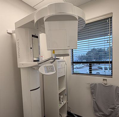
The scanning process itself is typically quick and noninvasive, often lasting less than a minute from the patient’s perspective. Patients are positioned comfortably and asked to remain still while the machine rotates. Unlike conventional medical CT scanners, dental CBCT units are designed to be compact and patient-friendly, minimizing the stress and time associated with imaging.
Before any scan, our clinicians review the indication for imaging and discuss what we hope to learn from the CBCT. This informed approach ensures that every scan has a clear diagnostic purpose and that patients understand the benefits and limitations of the study.
CBCT is most powerful when its information is integrated into a broader treatment strategy. The scans are reviewed alongside clinical exams, intraoral photos, and patient goals to create a plan that is individualized and realistic. Whether restoring a single tooth, planning full-arch rehabilitation, or evaluating oral pathology, CBCT contributes objective data that enhances decision-making.
Our practice uses CBCT findings to explain options to patients with clarity, showing relevant slices and reconstructions to illustrate anatomy and planned approaches. This visual information helps patients feel more informed about their care and fosters collaborative treatment planning between clinician and patient.
As imaging technology and software continue to evolve, the role of CBCT in dentistry grows as well. When used appropriately, it is a powerful adjunct that supports safer procedures, more predictable outcomes, and a higher standard of clinical care.
In summary, cone-beam computed tomography is a vital diagnostic tool that enhances accuracy across many areas of dental care. If you’d like more information about how CBCT might be used in your treatment or have questions about the process, please contact us to learn more.
Cone-beam computed tomography (CBCT) is a three-dimensional imaging technology that captures volumetric views of the teeth, jaws and surrounding structures. The scan produces distortion-free images that can be viewed in multiple planes, giving clinicians spatial information that conventional two-dimensional films cannot provide. Because it reveals relationships among teeth, bone and vital anatomy, CBCT supports more confident diagnoses and treatment planning for complex cases.
CBCT is used across many dental specialties when two-dimensional images lack sufficient detail. Typical indications include implant planning, assessment of pathology, evaluation of traumatic injuries, endodontic diagnosis and surgical mapping. By providing precise measurements and anatomical context, CBCT helps clinicians tailor procedures to each patient’s unique anatomy.
Traditional dental x-rays capture flat images that compress three-dimensional structures into a single plane, which can obscure depth and overlapping anatomy. CBCT acquires a volumetric dataset that can be sliced and reconstructed to show cross-sections, axial views and three-dimensional renderings for clearer spatial orientation. This difference is especially important when evaluating root morphology, bone contours or proximity to nerves and sinuses.
Another key difference is the ability to take targeted fields of view with CBCT to focus on a region of interest while limiting exposure. Image resolution and the type of diagnostic information available also vary between modalities, so clinicians choose the tool best suited to the specific clinical question. Both technologies remain valuable; CBCT complements rather than replaces conventional radiography in routine care.
CBCT is essential for accurate implant planning because it provides three-dimensional measurements of bone height, width and density. Clinicians can evaluate the available bone, identify critical structures such as the inferior alveolar nerve and maxillary sinus, and determine ideal implant angulation and position before surgery. This preoperative insight reduces the risk of unexpected findings during the procedure and supports safer, more predictable outcomes.
When bone grafting or sinus augmentation is being considered, CBCT helps quantify the volume of augmentation required and pinpoint the optimal graft locations. The data can also be used to design guided surgical templates that transfer the virtual plan to the clinical setting, improving the precision of implant placement. Postoperative scans provide a reliable baseline for monitoring healing when clinically indicated.
In endodontics, CBCT is particularly valuable for identifying complex root canal anatomy, locating additional or hidden canals, and detecting periapical pathology or vertical root fractures that may be missed on conventional films. Three-dimensional views help clinicians plan retreatments and assess the true extent of lesions. The technology guides more targeted interventions by revealing details of the root and surrounding bone.
Orthodontists use CBCT in complex cases that require a full understanding of skeletal relationships, impacted teeth or airway considerations. For TMJ assessment, CBCT provides clear visualization of osseous components such as condylar morphology and joint spaces, which aids in diagnosing bony degenerative changes. While soft-tissue evaluation still relies on other modalities, CBCT contributes important bony information to a comprehensive assessment.
Modern CBCT systems are designed to minimize radiation exposure while delivering diagnostically useful images, and clinicians follow the principle of keeping dose as low as reasonably achievable (ALARA). Exposure is controlled by choosing the smallest practical field of view and an appropriate resolution setting that matches the diagnostic task. This approach ensures scans are obtained only when the additional three-dimensional information will influence clinical decisions.
Before ordering a CBCT scan, clinicians assess the indication and discuss the benefits and risks with the patient to ensure informed decision-making. For routine, low-risk problems, conventional radiographs often suffice and are preferred; CBCT is reserved for situations where its 3D information changes diagnosis or treatment planning. Pregnant patients and others with specific concerns are evaluated on a case-by-case basis to determine the safest approach.
The CBCT scanning process is typically quick, noninvasive and well tolerated by most patients. After being positioned in the unit, the patient is asked to remain still while the scanner rotates around the head, and the actual acquisition usually takes less than a minute. Technicians provide guidance and ensure the patient is comfortable and stable before imaging begins.
There is no need for contrast agents or sedation in ordinary dental CBCT exams, and recovery time is not required because the procedure involves only passive imaging. Once the scan is complete, the dataset is reconstructed and reviewed by the clinician to determine next steps, and relevant images or slices can be shown to the patient to explain findings and treatment options.
CBCT provides excellent bony detail but has limited capacity for soft-tissue contrast compared with medical CT or MRI, so it is not the ideal modality when soft-tissue pathology is the primary concern. Image quality can also be affected by patient movement, metallic restorations or artifacts that may obscure fine details in certain regions. Clinicians weigh these limitations against the expected diagnostic benefit when deciding to image.
Contraindications are generally related to pregnancy or situations where alternative imaging would provide the needed information with lower risk. Younger patients are imaged judiciously to avoid unnecessary exposure, and protocols are adjusted to reduce dose when applicable. The decision to use CBCT always follows a clinical assessment and a clear diagnostic purpose.
Interpreting a CBCT scan involves reviewing multiplanar slices and three-dimensional reconstructions to identify anatomic landmarks, pathology and spatial relationships relevant to the clinical question. Clinicians correlate image findings with clinical exams, intraoral photos and patient goals to form a comprehensive diagnosis. This integrated review yields objective data that informs choices such as implant size and position, surgical approaches or endodontic strategies.
The CBCT data can also be exported to planning software for virtual simulations, measurements and fabrication of surgical guides when indicated. Sharing annotated images with patients helps explain recommended procedures and anticipated outcomes in a visual, patient-friendly way. When appropriate, the images are documented in the record and used as a baseline for follow-up evaluations.
Yes, CBCT datasets are commonly integrated into digital workflows to design and fabricate surgical guides, provisional restorations and prosthetic components. By combining CBCT scans with intraoral scans or digital impressions, clinicians and laboratory partners can plan implant positions virtually and translate that plan into a precise, guided surgical procedure. This coordination reduces intraoperative guesswork and improves the accuracy of implant placement.
Digital workflows that incorporate CBCT also streamline communication among the clinical team and dental laboratories, supporting predictable restorative outcomes. When a guided approach is selected, the practice follows verification protocols to ensure guide fit and surgical conditions match the virtual plan before proceeding with treatment.
At our practice, clinicians reserve CBCT for cases where three-dimensional information will materially affect diagnosis or treatment planning. The decision follows a thorough clinical evaluation that considers patient symptoms, clinical findings and the limitations of two-dimensional imaging. Imaging protocols are selected to match the diagnostic need while minimizing exposure through field-of-view and resolution choices.
When a CBCT scan is recommended, the clinician explains the specific reason for imaging, what the team hopes to learn from the scan and how the information will influence next steps. This patient-centered approach ensures that CBCT is used responsibly as a tool to enhance safety, predictability and the overall quality of care.